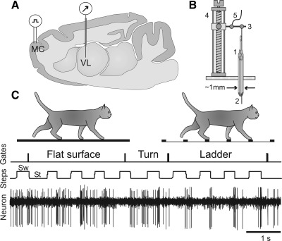
Fig. 1.
Experimental paradigm. A: schematic drawing of a parasagittal section of the brain showing the position of chronically implanted guide tubes for recording electrodes above the ventrolateral thalamus (VL) and stimulating electrodes in the forelimb representation of the motor cortex (MC). B: method of insertion and advancement of electrodes into the VL. 1: A group of 28-gauge cannulas is chronically implanted in the cortex above VL. 2: An electrode is manually inserted into one of the cannulas and soldered to an arm (3) of a micromanipulator (4). A wire (5) that leads to a miniature preamplifier positioned on the head of the animal is also soldered to the arm. In this manually driven micromanipulator, 1 revolution of the screw results in 200-μm advancement of the electrode. C: locomotion tasks: walking in a chamber on a flat surface and along a horizontal ladder. The Gates trace indicates when the cat has passed the beginning and end of each of the chamber's corridors. The Steps trace shows the swing (Sw) and stance (St) phases of the right forelimb recorded with an electro-mechanic sensor. The Neuron trace shows discharge of a neuron from the VL.

