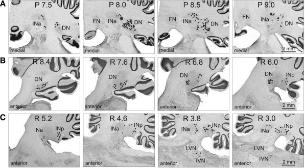
Fig. 3.
Cerebellar projections to the recording area in the VL. A: neurons in the anterior interposed (INa) and lateral (dentate, DN) nuclei in cat A, retrogradely labeled with WGA-HRP. Neurons are depicted with black circles on photomicrographs of coronal sections of the cerebellum contralateral to the injection site. B and C: neurons in the dentate, anterior, and posterior (INp) interposed nuclei and inferior vestibular nucleus (IVN) in cat B, retrogradely labeled with red fluorescent beads. Neurons are shown on photomicrographs of parasagittal sections of the cerebellum contralateral to the injection site. Each circle represents 1 labeled neuron. LVN, lateral vestibular nucleus.

