Fig. 5.
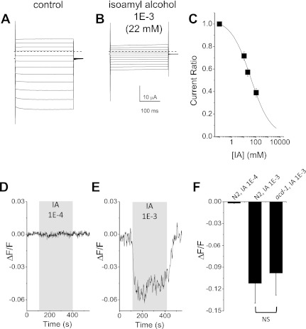
ACD-1 channels and glial amphid sheath cells are inhibited by high concentrations of isoamyl alcohol. A: family of ACD-1 currents in a Xenopus oocyte injected with acd-1 cRNA. Currents were elicited by voltage steps from −160 to + 60 mV in 20-mV increments in an oocyte perfused with a physiological solution. The holding voltage was −30 mV. B: the same oocyte shown in A was perfused with physiological solution plus 1E-3 isoamyl alcohol. C: isoamyl alcohol dose-response curve. Data are means ± SE; n = 17. Data were fitted by a sigmoid curve that indicates a Ki of 38 mM (1.7E-3 dilution). D: example of GCamP1.0 fluorescence that reports intracellular Ca2+ changes in amphid sheath cells of a wild-type animal (N2) during perfusion with 1E-4 isoamyl alcohol. At this concentration, there was no change in intracellular Ca2+ in glial amphid sheath cells. E: same as in D during perfusion of a wild-type animal (N2) with 1E-3 isoamyl alcohol. F: average fluorescence changes in amphid sheath cells of wild-type (N2) and acd-1 mutant animals perfused with isoamyl alcohol at 1E-4 and 1E-3 dilutions as indicated. Data are means ± SE; n = 8, 15, and 8, respectively. NS, not significantly different.

