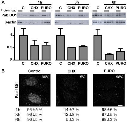
Figure 2. p53 decay upon translation inhibition does not correlate with Pab 1801 puncta disappearance.
U2OS cells were treated with cycloheximide (CHX) or puromycin (PURO) during 1, 3, or 6 hs. C, control. A, western blot with the Pab DO1. Duplicates of two-fold dilutions from each treatment were loaded. p53 levels were determined and normalized using beta-actin as loading control. Error bars, standard deviation. p53 decay was comparable upon protein synthesis inhibition by cycloheximide or puromycin. B, Cells were stained with the Pab 1801. Two representative cells upon 6 hs-treatment are shown. The percentage of cells with punctate Pab 1801 signal was evaluated in 100 cells from duplicate stainings for each treatment at the indicated time points. The Pab 1801 puncta vanished upon cycloheximide treatment and remained unaffected upon puromycin exposure. Bar, 10 µm.

