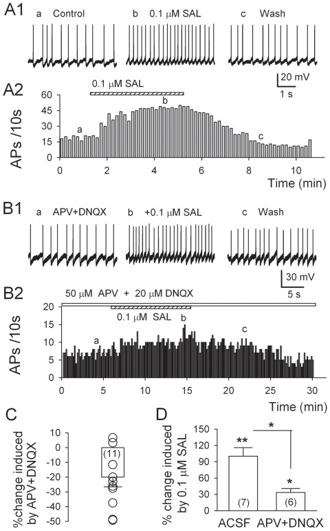
Figure 1. Salsolinol-induced stimulation of dopaminergic (DA) neurons is attenuated by APV and DNQX.
A1, Traces illustrate spike discharge at the times indicated in A2. A2, Time course of the increase in the ongoing pacemaking firing rate, recorded from a current-clamped DA neuron in the posterior ventral tegmental area (p-VTA) of a rat, by 0.1 µM salsolinol. B1, Traces obtained at the times indicated in B2. B2, Time course of the effect of salsolinol on the firing rate of a DA neuron in the presence of APV (50 µM) and DNQX (20 µM), the antagonists of NMDA and AMPA receptors. C, Summary plot of decease in firing rate by APV + DNQX. D, Summary plot (means ± S.E.M.) of increase in firing rate of p-VTA DA neurons induced by salsolinol (0.1 µM) in ACSF is larger than that in the APV+DNQX. Numbers in bars indicate numbers of neurons tested. *P<0.05, **P<0.01, paired t-test for salsolinol vs. pre-salsolinol control. Unpaired t-test for salsolinol vs. APV+DNQX+salsolinol.

