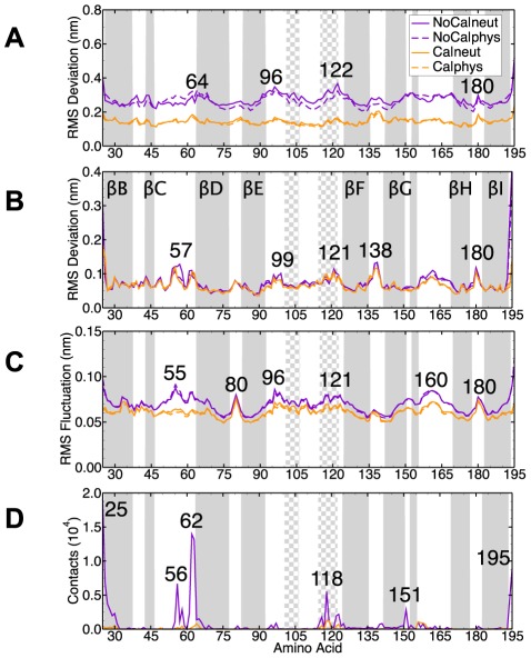
Figure 5. Structural dynamics of the capsid and water contacts.
(A) The RMSd from the crystal structure (without over all rotation and translation of the entire capsid) for the  of the shell domain (average over the last 0.1
  and all 60 proteins). (B) As A but after aligning each protein individually to the crystal structure. (C) The RMSf for the
  of the shell domain (average over the last 0.1
  ). (D) The number of contacts to water molecules crossing the capsid shell. The secondary structure elements of the crystal structure are shown as gray (
 -strand) or checkered (
 -helix) regions.

