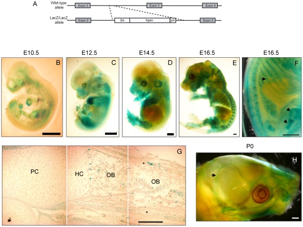
Figure 1. Prdm5 is expressed in osteoblast regions of developing bones.
A) Scheme for the generation of the Prdm5LacZ/LacZ mouse strain. B–E) X-gal stainings of Prdm5LacZ/LacZ embryos at E10.5 (B), E12.5 (C) E14.5 (D) and E16.5 (E). F) E16.5 embryo image detail. LacZ reporter expression in the perichondrium and growth plate of femur and ribs is marked by arrows. G) X-gal staining of tibiae section from E16.5 Prdm5 mutant embryo. Juxtaposition of three pictures (separated by white lines) to represent the whole length of a tibia. Indicated are different compartments: PC = proliferative chondrocytes, HC = hypertrophic chondrocytes, OB = osteoblasts. Periosteum is marked by asterisks. H) Whole mount X-gal staining of Prdm5LacZ/LacZ newborn skull at P0. Pronounced staining in sutures is indicated with an arrow. Bars = 1 mm, except for (G) where bar = 200 µm.

