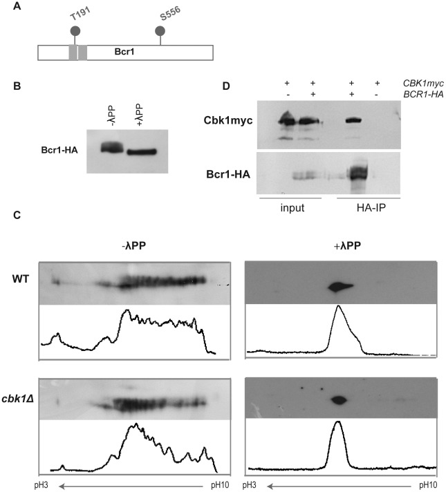
Figure 2. Bcr1 is a phosphoprotein that interacts in vivo with Cbk1.
(A) Schematic representation of Cbk1 consensus phosphorylation sites in Bcr1. Gray rectangles: Zn finger domains. (B) Cell extracts from a BCR1-HA strain (JC1144) grown at 37°C in Spider medium were analyzed by Western blot using anti-HA antibodies. A fraction from the same lysates was treated with λ-phosphatase (λPP). (C) Cell lysates from wild-type (WT, JC1144) and cbk1Δ (JC1159) strains grown in Spider medium at 37°C were subjected to 2D-WB and probed with anti-HA antibodies. Half of each lysate was treated with λ-phosphatase. Histograms obtained using ImageJ show the quantification of the relative intensity of each spot of the blot. (D) Protein extracts from a CBK1-myc BCR1-HA strain (JC1151) were immunoprecipitated using anti-HA antibodies. A strain carrying CBK1-myc (JC305) was used as negative control of the immunoprecipitation. Samples were separated by SDS-PAGE and probed with anti-myc or anti-HA antibodies.

