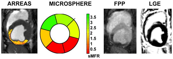
Figure 4.
Relation between ARREAS processed myocardial BOLD, segmental microsphere flow ratios bull’s eye plot, first-pass perfusion (FPP), and late-gadolinium enhancement (LGE) images. BOLD image (processed with the ARREAS method matched to the trigger time of the first-pass perfusion image), segmental microsphere flow ratios (sMFR) obtained by the ratio of flow between stress and rest for each segment, and first-pass perfusion image obtained under adenosine stress with critical LCX stenosis are shown for comparison. The late-gadolinium enhancement image acquired (at rest, prior to euthanization) at the same slice position and trigger time, confirms the absence of any infarction. Note the close correspondence between the BOLD image processed using ARREAS under the similar physiological conditions, the first-pass perfusion image and the microsphere bull’s eye plot.

