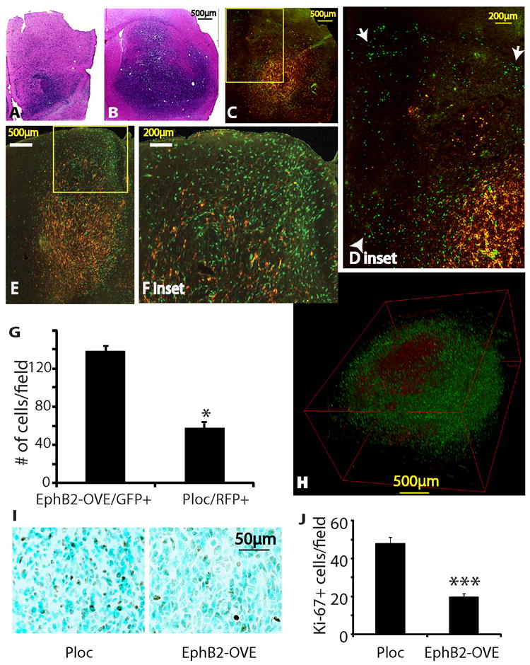
Figure 5. EphB2 controls the proliferation and migration dichotomy of GBM in vivo.

(A-F). Intracranial xenografts derived from the mixture of Ploc (5,000 cells, RFP+) and EphB2-OVE cells (5,000 cells, GFP+) indicated that EphB2 promotes tumor cell migration/invasion in vivo. Animals were sacrificed 67 days post-implantation. A, B. H&E stained sections of the invading margin (A) and the core (B) of a xenograft from the mixtures of Ploc and EphB2-OVE cells. C-F. Fluorescent images of two xenografts from the mixtures of Ploc and EphB2-OVE cells. The green EphB2-OVE cells were wide-spread, whereas the red Ploc cells mostly stayed in the tumor core, consistent with a more invasive phenotype of the EphB2-OVE cells in vivo. Enlarged images of the blocked area in C and E were shown in D and F, respectively. Arrow heads in D show cells invading into normal brain tissues and arrows show cells invading white matter tracts. (G). Quantification of EphB2-OVE cells (GFP+) and Ploc cells (RFP+) in the periphery of tumors indicated that there were 3-fold more EphB2-OVE cells than Ploc cells in the tumor periphery (***: P<0.05, n=3). (H). A three-D view of an intracranial tumor reconstructed from 40 adjacent brain sections. (I). Ploc (10,000) or EphB2-OVE cells (10,000) were separately implanted into mouse brain. Animals were sacrificed 67 days after implantation and brain sections were subjected for immunohistochemistry. Ki-67 staining showed that xenografts from Ploc cells had more cells under proliferation than that from EphB2-OVE cells. (J). Quantification of Ki-67+ cells in xenografts by taking pictures and counting positively stained cells. Xenografts from Ploc cells had 70% higher Ki-67+ staining than that from EphB2-OVE cells (*: P<0.05, n=10).
