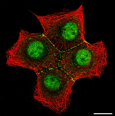
Fig. 3.
Localization of plakophilin-2 (Pkp2) in nuclei and desmosomes of cultured human breast adenocarcinoma-derived cells of line MCF-7. Double-label, confocal-laser scanning immunofluorescence microscopy showing Pkp2 (green) in a small colony of four MCF-7 cells, using polyclonal guinea pig Abs in comparison with the reactions of Abs against keratin IFs (red; murine mAb Lu-5). Note the specific staining of Pkp2 in the typical punctate arrays of desmosomal junctions and the dot-like exo- or endocytotic vesicles associated with desmosomal molecules in the cytoplasm, whereas the cell–cell junction free surface regions are negative. Note in addition the granular fluorescence staining of Pkp2 in the nucleus whereas the nucleoli are negative. Scale bar 20 μm

