Fig. 4.
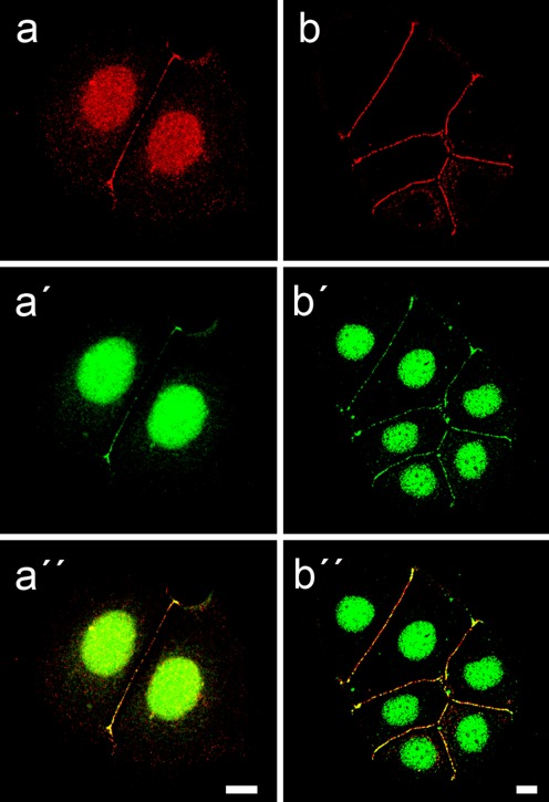
Specific dual localization of plakophilin-2 (Pkp2) in nuclei and desmosomes of cultured human breast adenocarcinoma-derived MCF-7 cells. a–a″ Confocal laser-scanning immunofluorescence microscopy of a double-label experiment, comparing the reactions of two Abs against different epitopes of Pkp2 (a, red: mAb Pkp2, clone Pkp2-518; a′, green: guinea pig Abs of serum GP-PP2) in two adjacent MCF-7 cells. The corresponding merged picture (a″) shows the colocalization of both kinds of Pkp2 Abs (yellow merged color) in desmosomes at cell-cell borders and in small granules in the nucleus. b–b″ Double-label immunolocalization micrographs of small colonies of MCF-7 cells, showing the frequent colocalization of Pkp2 (b′, b″; green) with the desmosomal plaque protein desmoplakin (b, b″; red). Note also the intense reaction of Pkp2 in the nucleus, in contrast to the absence of desmoplakin, while both proteins colocalize (b″, yellow merged color) in most—but not all—desmosomes in the cell-cell contact regions. Scale bars 10 μm

