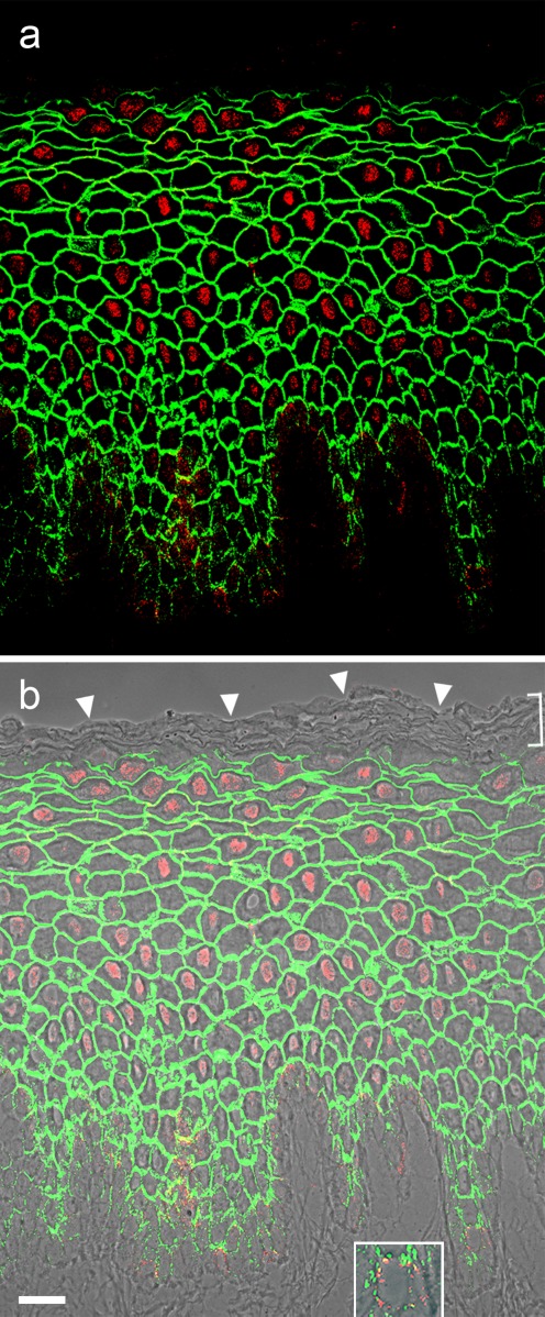
Fig. 5.
Nuclear localization of plakopilin-2 (Pkp2) in fetal porcine snout epithelium. a, b Immunofluorescence microscopy showing the localization of polyclonal guinea pig Abs specific for Pkp2 (red) on cryostat sections through fetal porcine snout epithelium in comparison with the desmosomal plaque protein desmoplakin (green). Under these immunostaining conditions used here the intense and specific nuclear localization of Pkp2 (red) contrasts with the desmoplakin immunostaining restricted to the desmosomes of all keratinocytes. Note also the absence of both to the uppermost layers of the stratum corneum (b, bracket; the tissue surface is denoted by the arrowheads). The picture of (b) is presented on a phase contrast background. Note in addition that Pkp2 is—in addition to the nuclei—also specifically located in distinct “dot-like” structures in the basal cell layer, representing one half of the heterotypic desmosomes connecting the keratinocytes and the neuroendocrine “Merkel cells” (insert in b shows a magnification of one of the Merkel cells in the basal layer in the right lower corner). Scale bar 20 μm

