Fig. 7.
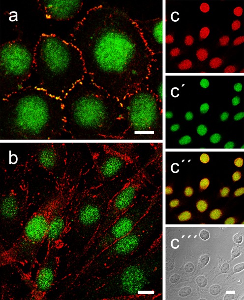
Differential localization of plakophilin-2 (Pkp2) in mammalian fibroblasts. Double-label, laser-scanning immunofluorescence microscopy of cultured transformed human fibroblasts of line SV80 (a), bovine dermal fibroblasts of line B1 (b) and mouse fibroblasts of strain L929 after formaldehyde fixation and detergent-treatment (for details, see “Materials and methods”). Here, the immunolocalization of polyclonal guinea pig Abs specific for Pkp2 (a, b, c′, c″; green) and the adherens junction (AJ) proteins N-cadherin (a, b; red) and β-catenin (c, c″; red) is shown. Under these conditions, both the specific staining of junctions at the cell–cell boundaries and the intense nuclear staining are seen. Note, however, that in the transformed fibroblasts (a), Pkp2 occurs in the N-cadherin-positive AJs (yellow merged color) whereas AJs of untransformed B1 fibroblasts lack Pkp2. In all fibroblasts, Pkp2 is also abundant in the nucleus (a–c) in comparable amounts as β-catenin as shown in the “junction-lacking” L929 fibroblasts (c–c″). The corresponding merged color picture (c″) and the phase contrast background (c″′) images are presented in addition in order to see the cytoplasms of these cells. Scale bars 10 μm

