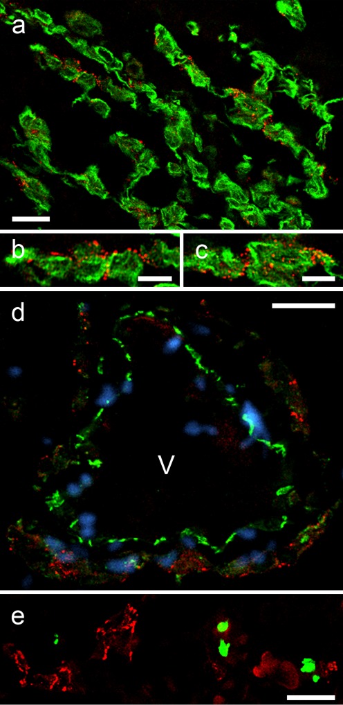
Fig. 9.
Localization of plakophilin-2 (Pkp2) in adherens junctions (AJs) of human cardiac myxomata. Laser-scanning, double-label immunofluorescence microscopy of sections through formaldehyde-fixed and paraffin-embedded human myxomata, treated for antigen-retrieval and double-immunostained with mAbs to Pkp2 (red), in comparison with green-labeled polyclonal Abs decorating IF bundles of the vimentin-type (a–c), the vascular endothelial transmembrane glycoprotein VE-cadherin (d) and the cell proliferation marker protein Ki67 (e). Note the extensive localization of Pkp2 in the AJs connecting myxoma cell processes, which also contain vimentin IFs (a–c). b, c Higher magnification micrographs of the region shown in (a), demonstrating that Pkp2-positive puncta adhaerentia are often clustered and appear as “beaded chains”. d Note here that the AJs connecting the endothelial cells of vessels (V), which are recognized by their intense VE-cadherin staining, are negative for Pkp2. Moreover, in myxomata, only a few cells undergo cell division as seen by immunostaining with Ki67 as proliferation marker (e). DAPI staining (blue) was used to visualize the nuclei in (d). V vessel lumen. Scale bars 10 μm (b, c), 20 μm (a, d, e)

