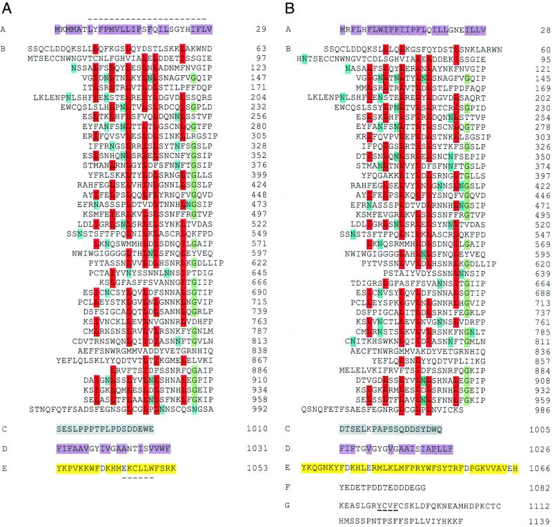
Figure 3.
Primary structure of the Ve1 (A) and Ve2 (B) proteins deduced from cDNA sequence. The polypeptides have been divided into domains A–G as described in the text. A dashed line occurs above the putative N-terminal leucine zipper in domain A of Ve1 and below the endocytosis signals in domain E of Ve1 and domain G of Ve2. Highlighted are the hydrophobic amino acids (purple) of the putative signal peptide domain A and membrane-associated domain D; conserved L/I (red), G (green), and potential N-glycosylation sites (blue) within the LRR domain B; neutral and acidic amino acids (gray) of domain C; and neutral and basic amino acids (yellow) of domain E. The PEST sequence of Ve2 is shown in domain F.

