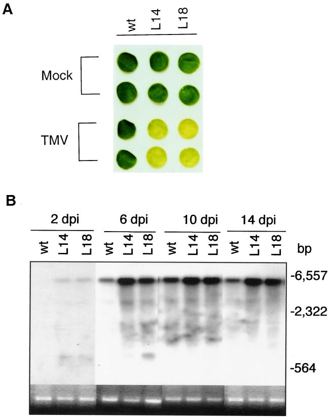
Figure 4.
TMV symptom development and viral RNA accumulation in detached leaf discs. (A) Leaf discs from wild-type or antisense lines 14 (L14) and 18 (L18) were inoculated with TMV. The inoculated leaves were incubated in Petri dishes for 14 days before photographing. The enhanced chlorotic symptoms typically began in the antisense lines 10 days after inoculation. (B) Total RNA was isolated from TMV-infected leaf discs at indicated days postinoculation (dpi), separated on an agarose (1.2%)-formaldehyde gel, and probed with a DNA fragment corresponding to the TMV coat protein subgenomic RNA. The blot for the 2-dpi time point was exposed twice longer to detect low levels of TMV RNAs. The ethidium bromide stain of rRNA is shown for each lane. Migration positions of size markers (generated from HindIII-digested phage λDNA fragments) are indicated.

