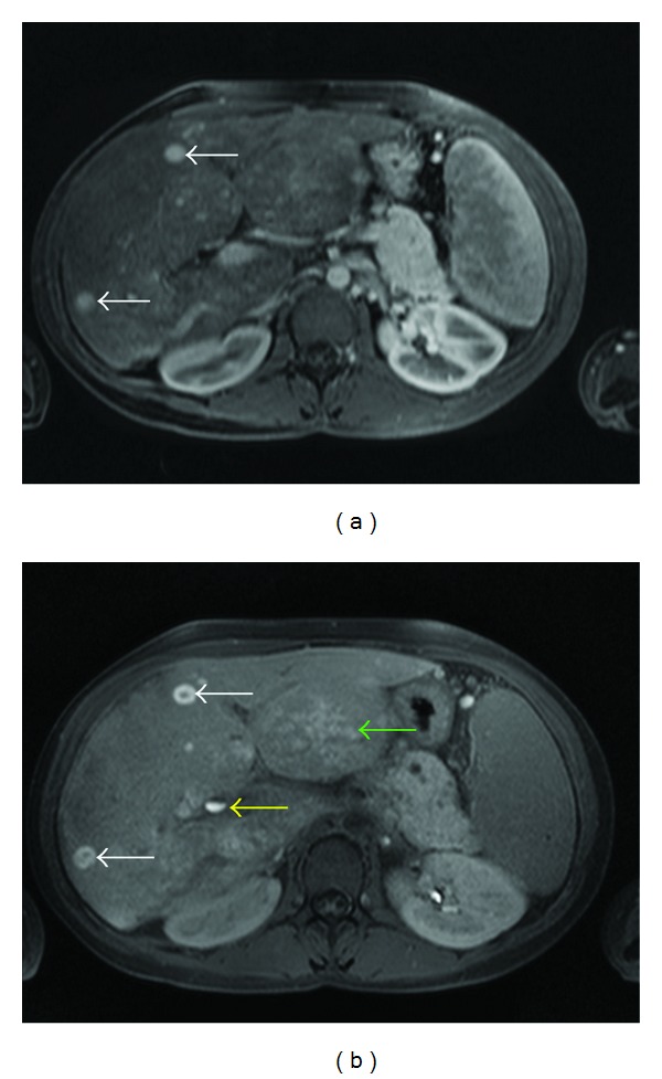
Figure 2.

As the multiple lesions show a homogeneous enhancement in portal venous phase (a) (T1-weighted VIBE sequence), there is a central “washout” with a peripheral pooling of contrast agent (white arrow) in late phase (b) (T1-weighted FLASH sequence, 20 min p.i.). This is likely to be due to a missing washin of the more central parts. Additionally, there is an evidence of an inhomogeneous hilar pooling of the contrast agent, predominantly in the left liver lobe (green arrow). Contrast agent level in the ductus hepatocholedochus (yellow arrow).
