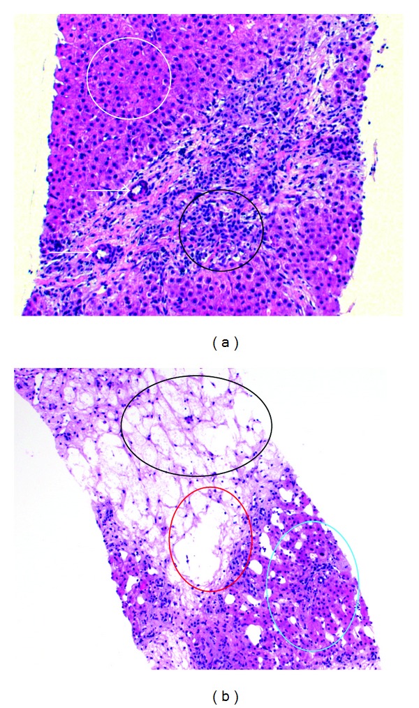
Figure 4.

Histological preparation: (a) thickened arterial vascular wall (arrows), proliferation of bile ducts and of inflammatory cells (black), and normal hepatocytes/liver parenchyma (white) (b) dropout of hepatocytes as a sign of cirrhosis of the liver, liver vein (red), and normal liver parenchyma (light blue).
