Figure 1.

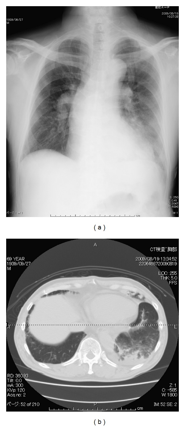
Chest X-ray and representative computed tomography image obtained on day 1. The patient had localized consolidation on his left lower lobe, suggesting postsurgical pulmonary edema in the dilated segment.

Chest X-ray and representative computed tomography image obtained on day 1. The patient had localized consolidation on his left lower lobe, suggesting postsurgical pulmonary edema in the dilated segment.