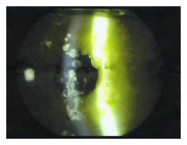
Figure 2.

A digitalized picture of the right eye, taken before LASIK was performed, shows the presence of multiple crumb-like and lattice-like stromal opacities consistent with the diagnosis of ACD. No granular infiltrates were present before surgery.
