Figure 2.

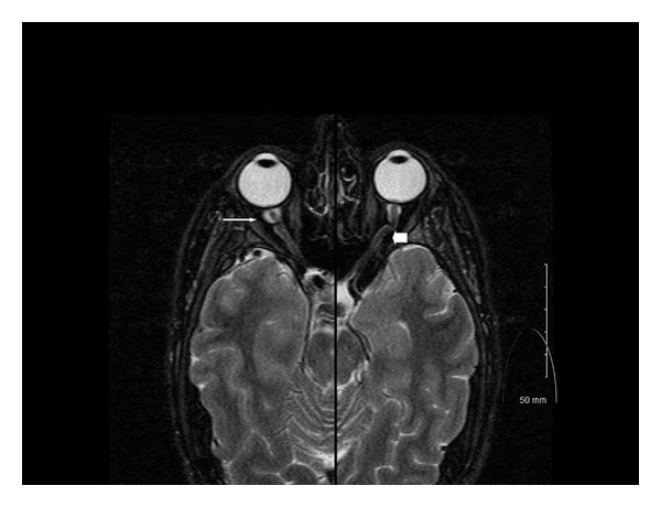
MRI of optic nerves. Merged axial T2 MRI at the level of the optic nerves shows significantly increased optic nerve sheath expansion near the globe (thin arrow), which extends through the course of the optic nerve sheath (thick arrow).

MRI of optic nerves. Merged axial T2 MRI at the level of the optic nerves shows significantly increased optic nerve sheath expansion near the globe (thin arrow), which extends through the course of the optic nerve sheath (thick arrow).