
Fig 4.
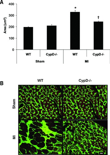
Cardiomyocyte cross-sectional area in remote myocardium of CypD–/– and WT mice 28 days after MI. (A) In WT mice but not CypD–/– mice, cardiomyocyte cross-sectional area increased significantly following MI, such that post-MI cross-sectional area was significantly greater in WT mice when compared to CypD–/– ones. (B) Representative high power photomicrographs of wheat germ agglutinin-stained LV cross-sections of non-infarcted myocardium. Bar = 10 μm. n= 7–10. *P < 0.05 versus respective WT sham. †P < 0.05 versus WT MI.