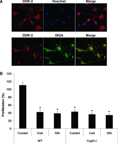
Fig 6.

Proliferation of cardiac fibroblasts isolated from WT and CypD–/– hearts. (A) Representative confocal micrographs of DDR2-stained (red) cardiac fibroblasts isolated from WT mice. Cells were counterstained with Hoechst-33258 (blue) for nuclei or wheat germ agglutinin (green) for cell membranes. (B) The proliferation of cardiac fibroblasts isolated from WT mice was significantly increased in response to 10% serum when compared to those isolated from CypD–/– mice. This increased in proliferation observed in the WT cardiac fibroblasts was prevented by CsA (0.4 μM) or SfA (1.0 μM). n= 4. *P < 0.05 versus WT control.
