Figure 3.
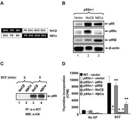
Cooperation between carboxy- and amino-terminal sequences of p85 is required for mast cell growth. (A) Schematic of p85 chimera constructs consisting of N-terminal p85α and C-terminal p85β sequences (NαCβ) or N-terminal p85β and C-terminal p85α sequences (NβCα). (B) P85α−/− mast cell progenitors (MCp) transduced with indicated p85 constructs were sorted to homogeneity and cultured in the presence of IL-3 (10 ng/mL). To detect the expression of p85 subunits, cells were harvested and subjected to Western blot analysis using anti-p85 PAN, anti-p85α, anti-p85β, and β-actin antibodies. (C) Murine myeloid 32D cells were coinfected with WT KIT and p85 chimera mutants (NαCβ or NβCα) and starved for 8 hours in serum- and cytokine-free media followed by SCF stimulation (100 ng/mL) for 5 minutes. An equal amount of protein (500 μg) was subjected to immunoprecipitation using an anti-KIT antibody followed by Western blotting with an anti-HA antibody. (D) WT and P85α−/− BMMCs transduced with vector or p85 constructs were sorted to homogeneity and cultured in the presence of IL-3 (10 ng/mL). Cells were starved for 6 hours in serum- and cytokine-free media and cultured in the presence or absence of SCF (50 ng/mL). After 48 hours, proliferation was evaluated by [3H]thymidine incorporation. Bars represent the mean [3H]thymidine incorporation in BMMCs (CPM + SD) from one representative experiment performed in quadruplicate. Similar results were observed in 6 independent experiments. *P < .01, WT-vector versus P85α−/−-vector versus P85α−/−-p85β versus P85α−/− -NαCβ. **P < .01, P85α−/−-vector versus P85α−/−-p85α versus P85α−/− -NβCα.

