Figure 7.

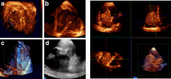
Left panel showing different examples of intracardiac thrombi into LV (A, B), RV(C), and LA appendage (D). Right panel showing quadscreen of RA myxoma.

Left panel showing different examples of intracardiac thrombi into LV (A, B), RV(C), and LA appendage (D). Right panel showing quadscreen of RA myxoma.