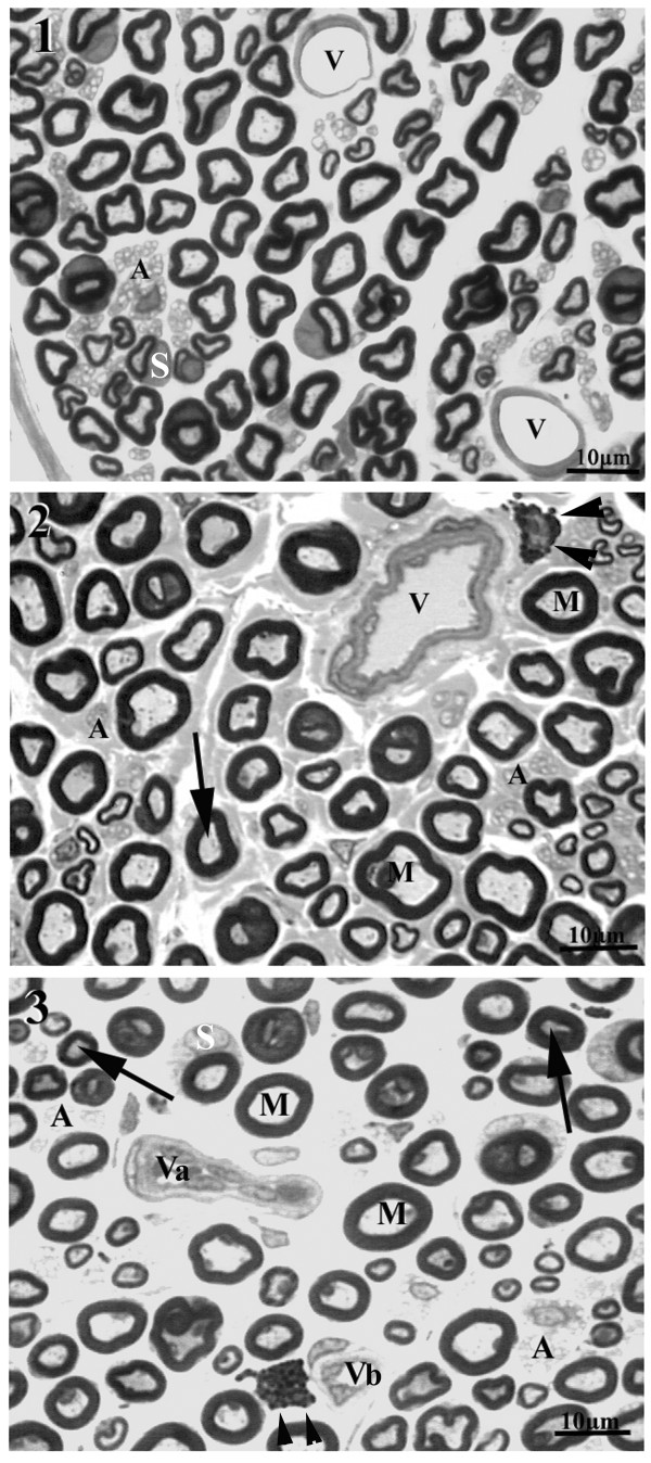
Figure 1.
Representative semithin cross section of the sural nerve of young (20 weeks old) male WKY (1) and male SHR (2 and 3), showing typical endoneural structures. Large (M) and small myelinated fibers as unmyelinated (A) fibers are present in the endoneural space. Mast cells are indicated by arrowheads and Schwann cell nuclei are indicated by S. Note the presence of normal endoneural vessels (V) in the WKY nerve while in SHR thickening of the wall (in 2), collapsed vessels (Va) and vessels with endoneural hyperplasia (Vb) were common. Arrows indicate large axons with atrophy. Toluidine blue stained. Bar = 10 μm.

