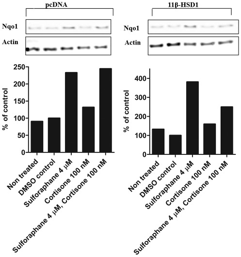
Figure 6. Suppression of NQO1 protein expression by cortisone in 11β-HSD1 expressing H4IIE cells but not in pCDNA3 transfected cells.
H4IIE cells transiently transfected with either pCDNA3 or 11β-HSD1 were treated for 24 h with vehicle (DMSO), cortisone, sulforaphane or cortisone and sulforaphane (upper panel). Cells were lysed, and equal protein amounts were used for Western blot analysis. Samples were probed for NQO1 using actin as a loading control. Lower panel, densitometric analysis of NQO1 bands normalized against b-actin. Graphs are representative of three independent experiments.

