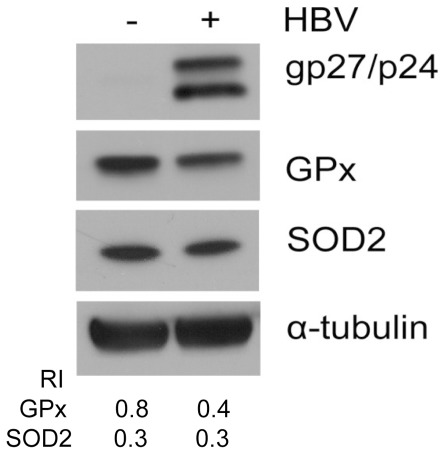
Figure 3. Suppression of GPx by HBV in C3A human hepatoma cells line.
C3A cells transiently transfected with pHBV1.3 or empty vector for 48 h were lysed for Western blot analysis for HBV small surface proteins (glycosylated/unglycosylated, gp27/p24), GPx and SOD2. α-tubulin was also analyzed to serve as a loading control. The RIs of GPx and SOD2 to α-tubulin are shown in the lower panel.

