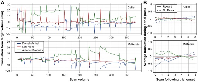
Figure 2. Motion during canine fMRI.
(A) Timeseries of translations required to correct for motion during the scan sessions. Volume 32 was the target for Callie, and volume 1 was the target for McKenzie. The plots therefore represent the total movement from the target volume. The spikes and breaks occurred when the dog moved its head out of the field of view, which typically happened following a reward. The volumes with artifacts were excluded from further analysis, leaving 62% of the volumes for Callie and 58% for McKenzie. Although the dogs did not place their heads back in exactly the same position, once they did, very little motion was observed. McKenzie exhibited a slow anterior-posterior drift during the second run, but this was sufficiently slow as to not cause movement artifacts during trials. (B) Average motion during a trial, separated by reward and no-reward conditions and after exclusion of volumes with artifacts. Scan volumes are 1610 ms apart. Notably, within-trial motion was less than 1 mm in all directions for both dogs, and no difference between the reward and no-reward conditions was observed.

