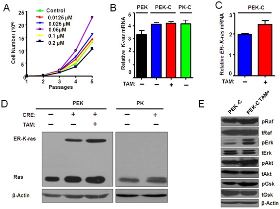
Figure 2. Treatment of tamoxifen at an optimal dose induced the activation of ER-K- rasG12D and downstream signaling.
A) The P53−/−, ER-K-rasG12D MEFs (PEK-C) derived from male embryo was counted for cell number after indicated dosage of tamoxifen treatment after 5 passages. B) Relative expression level of K-ras in indicated MEFs with or without tamoxifen treatment. PEK: P53L/L, Loxp-Stop-Loxp ER-K-rasG12D; PEK-C: P53−/−, ER-K-rasG12D; PK: P53L/L, Loxp-Stop-Loxp-K- rasG12D; PKC: P53−/−, K- rasG12D. (C) Detection of the ER-K-rasG12D expression in PEK-C cells with or without 0.05 µM tamoxifen treatment. D) Detection of K-ras and ER-K-ras protein level in MEFs infected with or without adeno-Cre in the presence or absence of 0.05 µM tamoxifen treatment in indicated MEFs. β-actin serves as internal control. E) The activation of Ras effector PI3K pathway signaling was confirmed by western blot.

