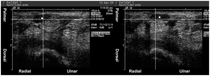
Figure 1. Example of median nerve motion direction measurement in middle finger motion in a patient.
The centroid of the median nerve (white dot) was taken in extension (left picture) and flexion (right picture) to calculate motion direction. The grid shows the change in position of the median nerve centroid in ulnar-palmar direction.

