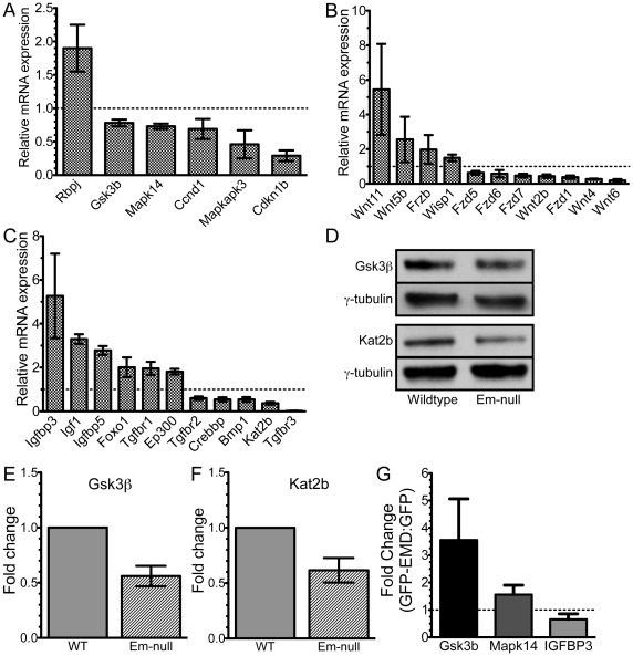
Figure 2. Validation of selected components of the Notch, Wnt, TGF-β and IGF pathways.
Validation of mRNA expression changes of selected Notch, Wnt, TGF-β and IGF pathways by qPCR. Dotted line represents no change in expression. A) Notch and p38 components. B) Wnt components. C) IGF and TGF-β components. All changes in gene expression were statistically significant (p≤0.05). D) Whole cell lysates of wildtype and emerin-null H2K myogenic progenitors were separated by SDS-PAGE and western-blotted for GSK3β, Kat2b and γ-tubulin. E,F) Densitometry was performed on blots in panel D and GSK3β and Kat2b levels were normalized to γ-tubulin. Emerin-null protein expression was normalized to protein expression in wildtype progenitors. G) GFP-emerin or GFP was expressed in emerin-null cells and Gsk3b, Mapk14, Igfbp3 and Gapdh were measured by qPCR. Data is depicted as fold-change in GFP-emerin expressing cells compared to GFP-transfected cells.

