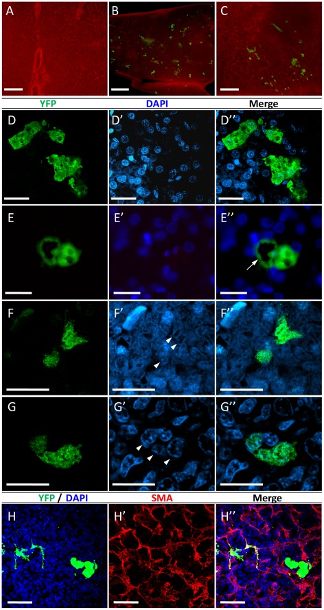
Figure 3. Immunofluorescence analyses of YFP expression in the liver and spleen of post-hatch chicks following in ovo FIV administration.
Whole-mount immunostaining with anti-GFP antibody (green) and anti-SMA antibody (red) was performed on pieces of livers from: 2-day-old chicks treated with either PBS (A) or FIV-YFP (B), and from 40-day-old FIV-YFP-treated chicks (C). Images were obtained using epi-fluorescent stereomicroscope. Scale bar = 200 µm for A and C, and 0.5 mm for B. Immunostaining of paraffin sections with anti-GFP antibody was performed on liver (D & E) and spleen (F & G) tissues from 2-day-old chicks treated with FIV-YFP. For each of these sections, DAPI staining (blue) is shown in the corresponding images (D′–G′) to indicate cell nuclei. Arrowheads mark nuclei of YFP-expressing cells with splenocyte morphology (F′). Merged YFP and DAPI staining is shown in the corresponding D′′–G′′. Images were obtained by confocal microscopy except for E, E′ and E′′, which were obtained by epi-fluorescenct microscope. The arrow in E′′ indicates a YFP-expressing cell with endothelial morphology located next to transduced cells with hepatocyte morphology. H. Confocal images of whole-mount immunostained liver sample, using both anti-GFP (H) and anti-SMA (H′) antibodies, confirmed the association of some of the YFP-expressing cells with blood vessels, which appear in yellow in the merged image (H′′). Scale bar = 20 µm (D-G) and 50 µm (H).

