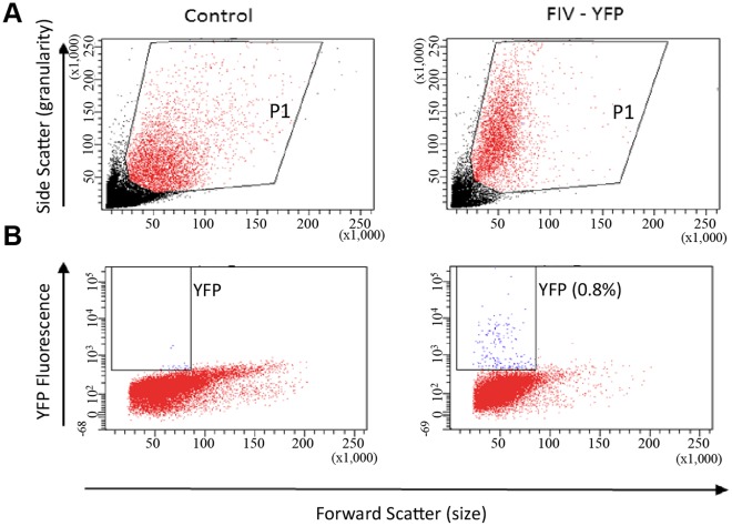
Figure 5. Estimation of transduction efficiency in livers of in ovo FIV-YFP transduced chicks by flow cytometry.
Liver samples from 2-day-old control chicks treated with PBS (A) or FIV-YFP (B) were analyzed by flow cytometry. One representative image from the control PBS and FIV-YFP treated chick groups is shown. Cells in each sample were selected (window P1 in A) and YFP fluorescence was analyzed (B). While no obvious signals were obtained in the control samples, the rate of transduced liver cells in the samples from FIV-YFP treated chicks was 0.46±0.19% (n = 3). Blue and red mark the YFP-positive and negative cells, respectively. The sample with the highest transduction rate (0.8%) is presented.

