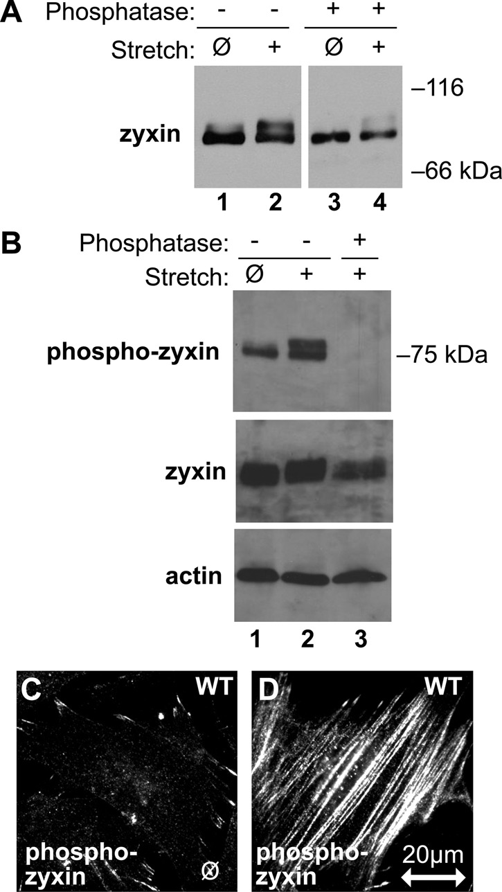
FIGURE 2:

Posttranslational modification of zyxin in response to stretch. (A) Immunoblot analysis of zyxin from unstretched (ø) and stretched (+) WT fibroblasts (lanes 1 and 2) identified a stretch-induced shift of zyxin, which was reversed after incubation with phosphatase (lanes 3 and 4). (B) Immunoblot analysis with a phosphospecific zyxin antibody on unstretched (ø) and stretched (+) cell lysates (lanes 1and 2) and elimination of the phospho-zyxin signal by incubation with phosphatase (lane 3). Immunoblots for total zyxin and β-actin show that their signals are retained following phosphatase treatment. (C, D) Indirect immunofluorescence microscopy on unstretched (ø) and stretched (+) fibroblasts with phospho-zyxin antibody. Phospho-zyxin signal is low at focal adhesions of unstretched cells, but it increases and appears along stress fibers in stretched cells. Stretch direction is in the horizontal plane (double-headed arrow; scale, 20 μm).
