FIGURE 7:
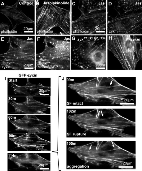
Actin stress fibers formed without VASP recruitment exhibit enhanced sensitivity to actin inhibitor jasplakinolide (Jas). (A, B) In comparison to controls, WT cells treated with jasplakinolide (100 nM, 2 h) accumulated stress fibers and aggregates of F-actin (phalloidin) visible by fluorescence microscopy. F-Actin (phalloidin; C) and zyxin (D) colocalized within the jasplakinolide-induced aggregates. Zyxin (E) and binding partner Mena/VASP (F) also colocalized at the jasplakinolide-induced aggregates. Jasplakinolide treatment (100 nM, 2 h) of zyxin-null cells expressing either GFP- zyxF71,93,105,115A mutant (G) or GFP-zyxin (H) induced a more pronounced actin phenotype, with disruption of zyxin-Mena/VASP interaction (zyxF71,93,105,115A). (I) Time-lapse microscopy of GFP-zyxin in cells (0, 30, 60, 90, and 114 min after 200 nM jasplakinolide addition) showed mobilization from focal adhesions to actin stress fibers, followed by rupture of fiber and accumulation of aggregates (J, arrows; 99, 102, 105 min). See Supplemental Video S1.

