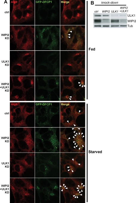
FIGURE 8:
Regulation of mAtg9 traffic by ULK1 and WIPI2. (A) Confocal microscopy of HEK293/GFP-DFCP1 cells treated with siRNAs against RISC-free (ctrl), ULK1, WIPI2, or WIPI2 and ULK1. Cells were incubated in full medium or starved for 2 h before fixation. mAtg9 was detected by indirect immunofluorescence. Arrows indicate areas of colocalization between GFP-DFCP1 and mAtg9. In WIPI2 KD virtually every DFCP1 spot colocalizes with mAtg9. (B) The efficiency of the knockdowns in A was confirmed by Western blot using antibodies against ULK1, WIPI2, and β-tubulin. One representative experiment of three is shown.

