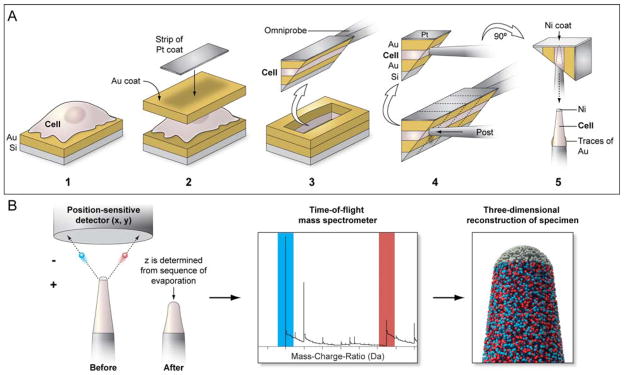
Figure 1. Schematic for the cellular sample preparation and APT data acquisition run.
(a) HeLa cells grown on gold-coated silicon substrates were coated with a thick layer of gold followed by a strip of platinum for further protection. Using FIB based protocols, a wedge containing a “sandwiched” cell volume was extracted and a sub-volume was attached to a microtip post in an orthogonal orientation. Following another protective nickel coat, the sub-volume was shaped into an APT-amenable tip by further FIB milling. (b) During an APT acquisition run, ions from the tip are extracted and are detected by a position-sensitive detector; this part of the tip is consumed as a result. Time-of-flight measurements of these detected ions allows their chemical identification in a mass spectrum, and a reverse-point algorithm can then be used to spatially place the ions in a high resolution reconstruction of the original tip.

