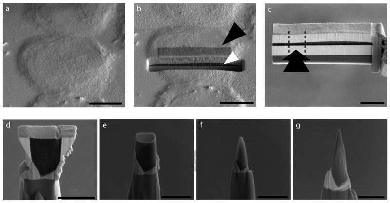
Figure 2. Biological sample preparation for APT.
(a) HeLa cells grown on gold-coated silicon substrates, flash-frozen and sputter-coated with gold. (b) A sub-area of a selected cell protected with a layer of platinum (black arrowhead); Focused Ion Beam (FIB) milling reveals the profile of the cell sandwiched between layers of gold (white arrowhead). (c) Cellular material after extraction as a wedge and attachment to an Omniprobe tip. The cell was attached to a post from the side (arrow) and separated from the rest of the cell by FIB milling (dotted lines). (d) Sub-volume of the HeLa cell stably attached to a probe tip. (e–g) Gradual sculpting of the sample into the needle-shape required for APT by annular FIB milling with decreasing radius. Scale Bars: a, b, 10μm; c, 5 μm; d–f, 2 μm; g, 1 μm.

