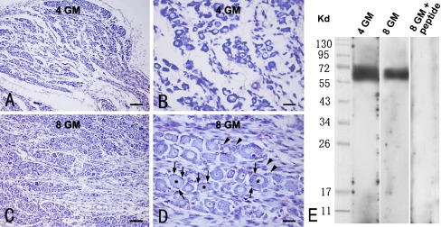
Fig. 1.
Histological evaluation of the prenatal human dorsal root ganglion (DRG) (a–d) and Western blot characterization of the rabbit anti-P2X3 antibody used in the present study. a–d Representative Nissl stain images of DRGs from 4 (a, b)- and 8 (c, d)-gestational month-old fetuses. The junction between the DRG and the peripheral spinal root is around the lower-right corner of the images (a, c). Nissl-stained neurons are round or oval with Nissl substance confined within the perikarya and the proximal neurites in some cases (b, d). Note that the cross-sectional area of the neurons appears larger in the 8-month (d) relative to the 4-month (b) samples. Nissl bodies are clearly seen in large DRG neurons in the 8-month case (d). Asterisks in d point to DRG neurons. Arrows point to satellite astrocytes (d). Arrowheads indicate other small DRG cells between neuronal islands (d). e The rabbit anti-P2X3 antibody blots a monomer band migrated at approximately 64 kd in DRG extracts from 4 (left lane) as well as 7 and 8 (mixed extract, middle lane) gestational month (GM) fetuses. This immunoblotting band is eliminated in the presence of the absorbing antigenic peptide in the primary antibody incubation buffer (right lane). Scale bar = 80 μm in a, c and 20 μm in b, d

