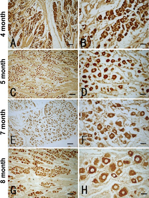
Fig. 2.
Representative images showing low (left panels) and high (right panel) power views of P2X3 immunoreactivity in the DRG from fetuses at four age points as indicated on the left. Immunoreactive cells are arranged as islands or clusters in all cases, with labeling occurring in the somata excluding the nucleus. Labeled perikarya are round, oval, or multipolar, sometimes with short proximal processes (B, D, F, H). Most labeled perikarya are less than 20 μm in diameter, but cells larger than 20 μm are increasingly seen in the 7- and 8-gestational month-old cases (compare D, F with B, D). The labeling intensity is heavy in most labeled cells in 4- and 5-month-old fetuses (B, D). The reactivity appears more differential in the DRGs of 7- and 8-month-old cases, wherein larger cells exhibiting lower intensity, with a granular appearance of the immunoreaction product in the cytoplasm (F, H). Note the immunoreactive fibers and bundles in A–F not in G, H. Scale bar = 80 μm in A, C and 20 μm in B, D

