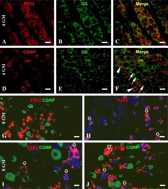
Fig. 5.
Representative double immunofluorescent images illustrating the cellular expression of P2X3 and CGRP in prenatal human DRG. a–f A 4-gestational month fetus and g–j an 8-month fetus. Neither P2X3 (a–c) nor CGRP (d–f) immunoreactive cells are colocalized with glutamine synthetase (GS). Arrows in f point to CGRP negative neurons, whereas arrowheads indicate CGRP-positive DRG neurons, both of which are surrounded by GS-labeled satellite astrocytes. Most P2X3-positive cells are not colocalized with CGRP (g, i), although coexpression of these two markers is seen occasionally (j, indicated by a triangle). Note that CGRP-labeled cells appear somewhat larger in somal size relative to P2X3 cells (g, i, j). In double and triple labeling preparations (h–j), P2X3 immunoreactive cells are frequently colocalized with IB4 binding (examples are indicated by arrows). Scale bar = 20 μm for a–h, j, and 10 μm in i

