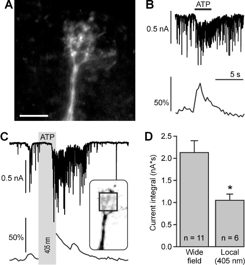
Fig. 4.
Photolysis of caged ATP evokes calcium signaling in the dendritic tuft of mitral cells. a Confocal image of a Fluo-8-filled dendritic tuft. Scale bar, 30 μm. b Synaptic currents (upper trace) and calcium signaling (lower trace) elicited by wide-field photolysis of NPE-ATP (100 μM). c Synaptic currents (upper trace) and calcium signaling (lower trace) elicited by local photolysis of NPE-ATP (100 μM). The calcium imaging was interrupted during the time of ATP photolysis (gray bar). The square in the inset, depicting the dendritic tuft, indicates the area where the 405-nm laser was directed to. d Local photolysis of caged ATP evoked significantly smaller currents compared to wide-field photolysis. *, p<0.05

