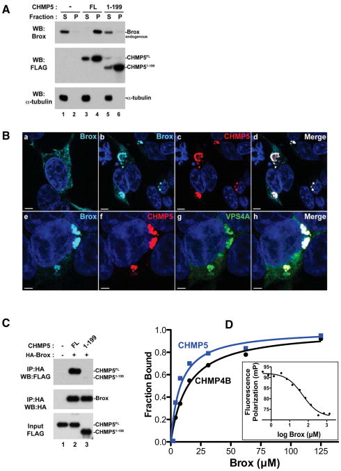
Figure 2. CHMP5 recruits Brox to VPS4A-positive endosomes and the CHMP5 C-tail is essential for Brox:CHMP5 interaction.
(A) CHMP5 recruits Brox to cellular membranes. HEK293T cells were transfected with empty vector (lanes 1–2), FLAG-tagged CHMP5 FL (lanes 3–4) or 1–199 (lanes 5–6). Forty-eight hours post transfection, cells were lysed and fractionated into soluble (S) and pellet (P) fractions as described in Experimental Procedures. Distribution of FLAG-tagged ESCRT-III and endogenous Brox was analyzed by western blot using the indicated antibodies. α-tubulin was used as a control protein for the soluble fraction.
(B) Brox and CHMP5 colocalize to VPS4A-positive endosomes. HA-tagged Brox was expressed in HEK293T cells alone (a), with FLAG-tagged CHMP5 (b–d) or in combination with both FLAG-tagged CHMP5 and GFP-tagged VPS4A (e–h). Cells were immunostained as described in Experimental Procedures. Brox, CHMP5, GFP-VPS4A are stained turquoise, red and green, respectively. Merge panels show colocalization of Brox and CHMP5 (d) or Brox, CHMP5 and VPS4A (h) in white. Scale bar = 5μm.
(C) The Brox binding site maps to the last 20 residues of CHMP5. FLAG-tagged CHMP5 full-length (FL, 1–219) or the C-terminal truncated fragment (1–199) was expressed in HEK293T cells in the presence or absence of the HA-tagged Brox. Examination of the protein interactions was performed similar to (A).
(D) Both CHMP4B and CHMP5 C-tails bind Brox. The C-tails of CHMP4B and CHMP5 bind Brox with dissociation constants of 14.3±1.4 μM (black) and 8.1±2.5 μM (blue), respectively, as measured by the FP assay. CHMP5 competes with CHMP4B for binding to Brox (insert) with an IC50 of 49.9 μM.
See also Figure S1.

