
Fig. 3. Runx2 colocalizes with G9a and enhances its intranuclear mobility.
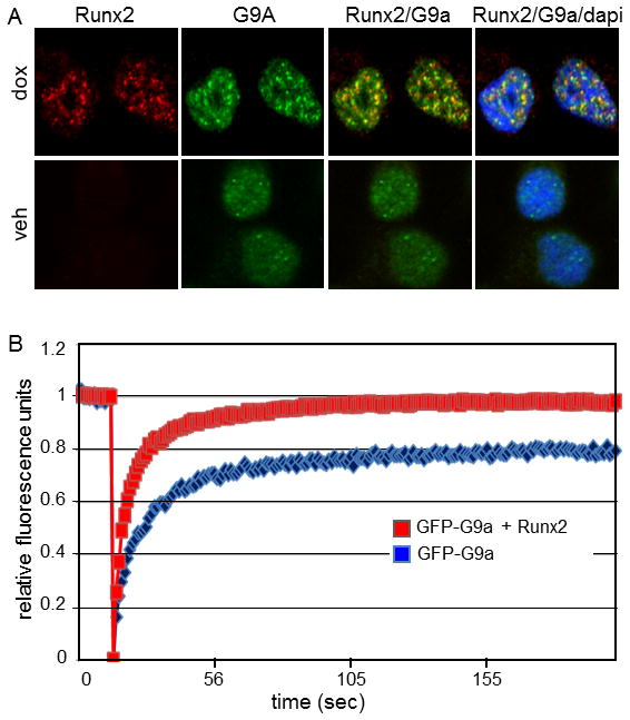
A: C4-2B/Rx2dox cells were treated for 24 h with dox to induce Runx2 expression or vehicle (veh) as control, and immunofluorescence analysis was performed to detect G9a (green) or Runx2 (red) proteins. DAPI staining (blue) indicates dense chromatin organization in the cell nuclei. Runx2/G9a, overlay of red and green images (yellow) indicates overlap; Runx2/G9a/dapi, overlay of red, green, and blue images. B: Fluorescence recovery after photobleaching of G9a-GFP was assessed as described in Materials and Methods in Cos7 cells transfected with plasmid encoding GFP-G9a alone (blue curve) or together with plasmid encoding Runx2 (red curve).