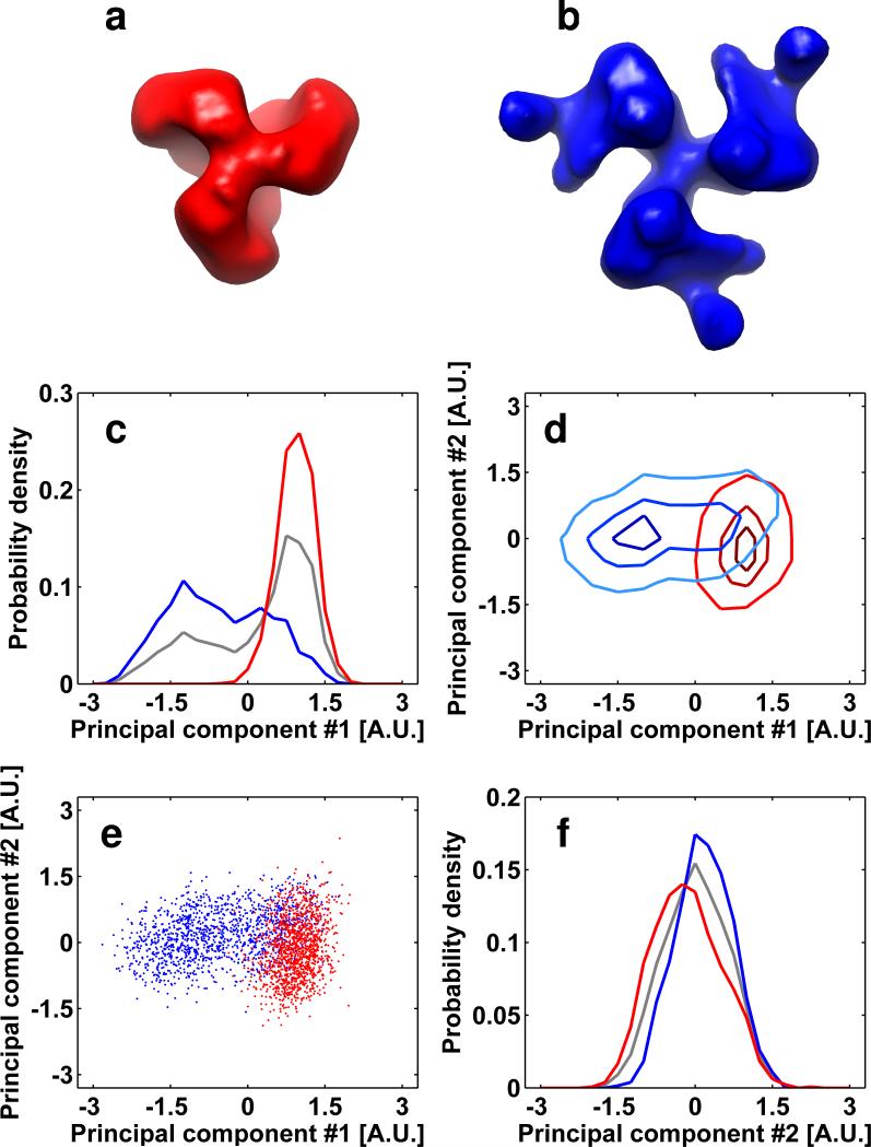
FIGURE 8.
Separation between soluble spikes in different conformations and ligand binding. Top views of the soluble spike in a closed unliganded state and in an opened sCD4 liganded sates are illustrated in panels a (red) and b (blue) respectively. (c-f) Visualization of the reduced dimensionality representation of the JR-FL SOSIP/JR-FL SOSIP+sCD4 mixture after iterative alignment and classification. Lines and markers related to unliganded and sCD4-liganded SOSIP trimers are colored red and blue, respectively. The gray lines in panels (c) and (f) are the normalized histograms of all of the data represented in the mixture. The overlap between the scatter-clouds of the sub-volumes representing unliganded and liganded complexes is only ~26%.

