Figure 4.
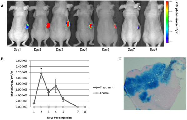
GLV-1h68-mediated luciferase and β-galactosidase expression in fibrosarcoma murine xenograft flank tumors. Fibrosarcoma (HT-1080) flank tumors were treated with a single dose of GLV-1h68 (1×10⁷ pfu) or saline as control. (A) Luciferase activity was measured after retro-orbital coelenterazine injections over a 8-day period. The reduction of luciferase activity correlated with tumor size regression. (B) Quantification of luciferase activity over a 8-day time course using software assessment of emitted protons was performed (n=3 mice). (C) HT-1080 flank tumors were excised 3 days after GLV-1h68 injection and stained for β-galactosidase expression. Representative photograph was taken by microscopy (10×). No animals demonstrated any evidence of toxicity.

