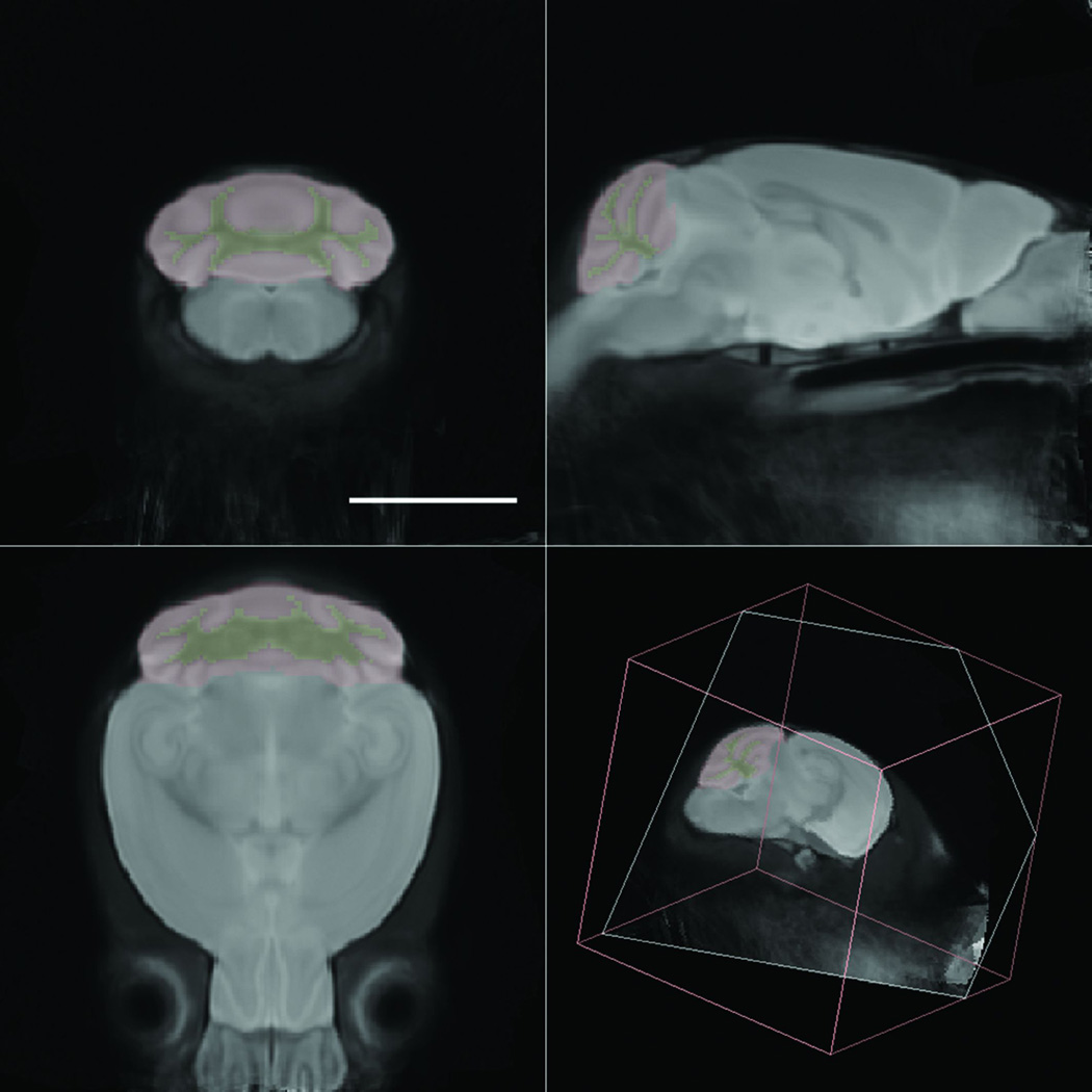
Fig. 2.
Minimum deformation atlas and delineations. A minimum deformation atlas was constructed from 30 postmortem high-resolution isotropic diffusion-weighted images of mouse brain. The anatomical delineations of the cerebellar cortex (red) and cerebellar white matter (yellow) are overlaid on the image. Scale bar = 5 mm.

