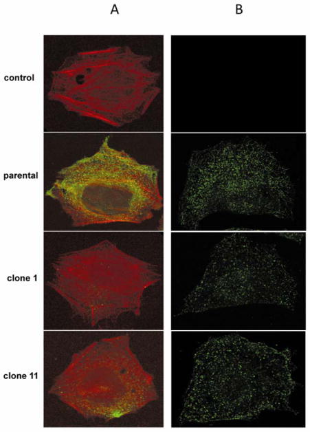
FIG. 3. Immunofluorescent staining for PDGFRα.
Parental and CHO FD11 Clones 1 and 11 were stained for PDGFRα expression. A) Cell surface PDGFRα expression was detected on unpermeabilized cells by staining with anti-PDGFRα (green) and counter staining with phalloidin (red). B) Intracellular PDGFRα was detected by permeabilizing the cells with Triton X-100 and then staining with anti-PDGFRα (green). Anti-PDGFRα was omitted in control samples in top row. Limited cell surface expression was detected in the CHO FD11 clone 1 and 11 cells compared with parental CHO K1 cells.

